Few biotechnology headlines are more tempting than the phrase “printed organs.” It suggests a future where a patient needs a kidney, a surgeon presses a button, and a perfect replacement arrives from a machine. The image is powerful because organ shortages are real, transplant medicine is extraordinary, and the idea of building spare parts for the body feels both humane and futuristic.
The reality is more interesting and less instant.
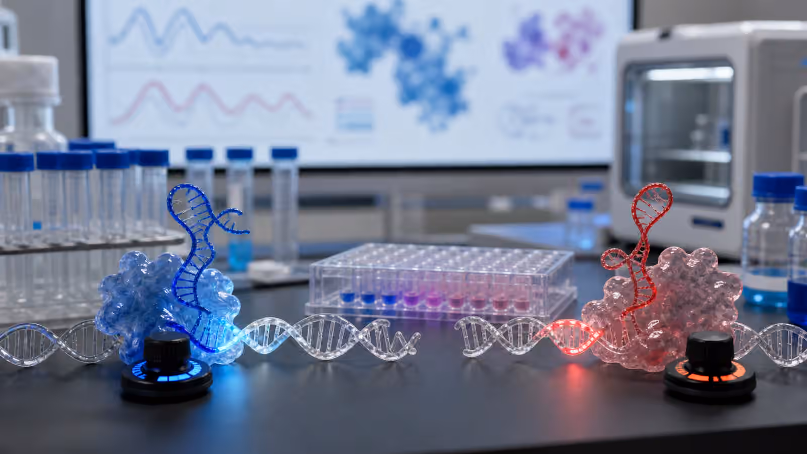
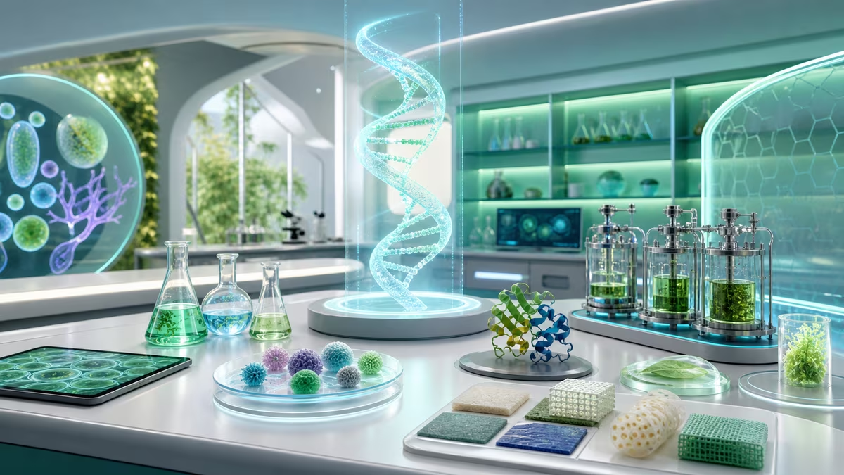
Tissue printing, often called bioprinting, uses cells, biomaterials, and printing-like methods to place biological material in organized patterns. It can create small tissue models, layered structures, scaffolds seeded with cells, and experimental constructs that help researchers study disease, test drugs, or explore regenerative medicine. It is part of the broader world of biofabrication .
But printing a living organ is not like printing a plastic phone case. Organs are vascularized, innervated, mechanically active, immune-interacting, self-repairing, metabolically demanding structures. They are not simply shaped tissue. They are living systems integrated with a body.
What tissue printing can do now
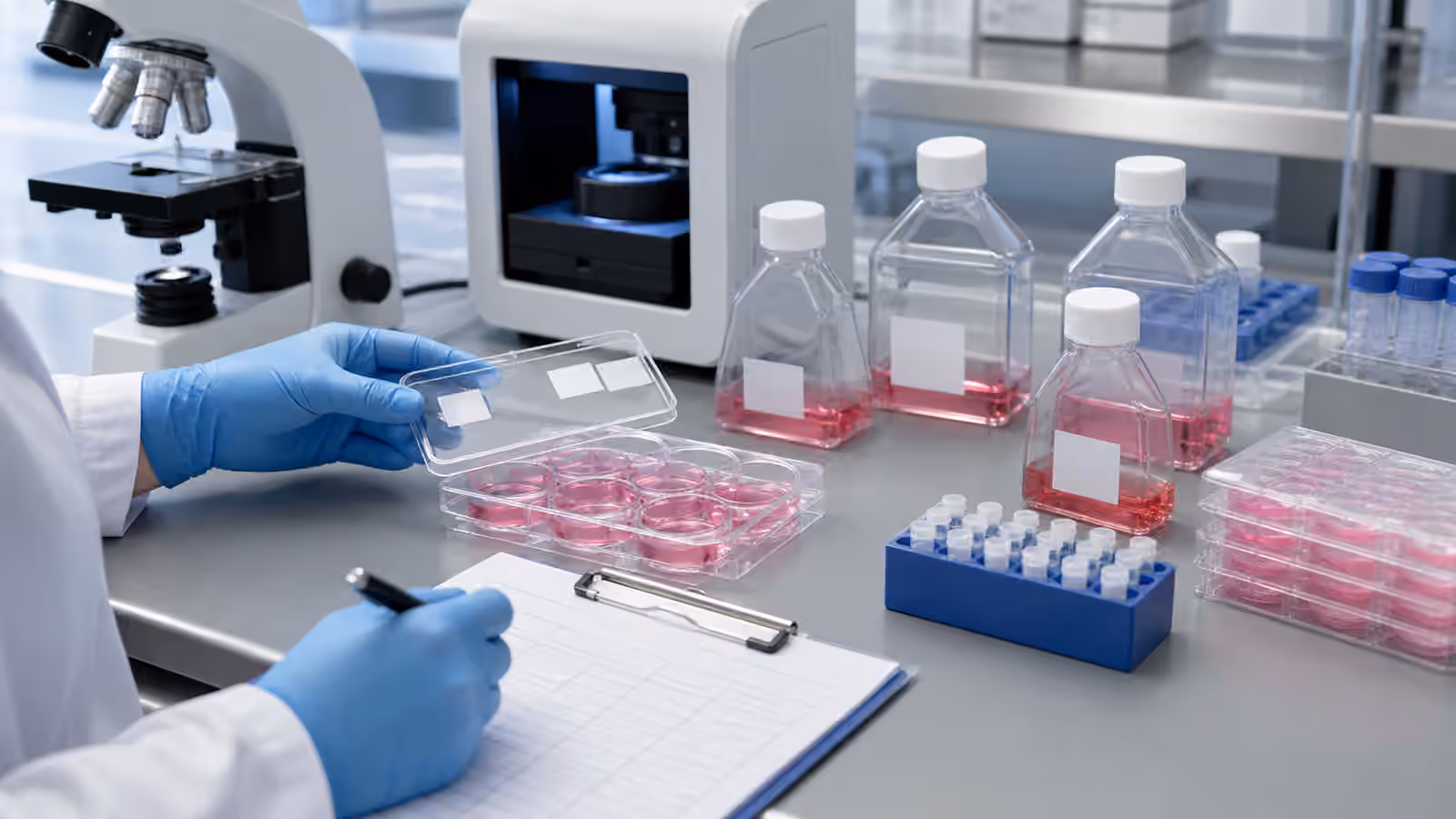
The most grounded uses are small and valuable. Researchers can print or assemble tissue-like models that mimic aspects of skin, liver, cartilage, blood vessels, tumors, or other tissues. These models can help study how cells behave in three dimensions, how drugs affect human-like tissue, or how disease changes structure.
A tissue model does not need to be a full organ to be useful. A tiny liver-like model that reveals toxicity can improve drug development. A skin model can help test irritation or wound-healing ideas. A tumor model can help researchers study how cancer cells interact with their surroundings. A vascular channel model can help study blood vessel behavior.
In medicine, partial tissues and scaffolds may support repair. Cartilage, bone-like structures, skin substitutes, and wound-healing materials are more plausible near-term targets than complex organs. They still require careful testing, but their demands are narrower than a kidney or heart.
How bioprinting works in plain English
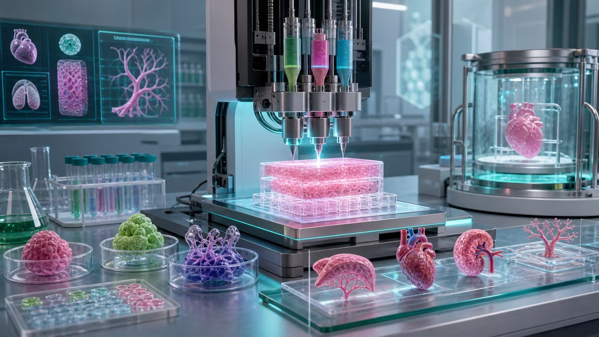
A normal 3D printer deposits plastic, resin, metal powder, or another material according to a digital design. A bioprinter may deposit “bioink,” a mixture that can include living cells, gels, proteins, polymers, nutrients, or support materials. The printer places material in layers or patterns. After printing, the construct must mature. Cells need time to attach, communicate, organize, remodel their environment, and sometimes differentiate.
The printing step is only the beginning. A printed tissue must stay alive. Cells need oxygen and nutrients. Waste must leave. Mechanical forces matter. The material must be soft or stiff in the right way. The cells must be the right type and behave appropriately. The construct must avoid contamination. If it is intended for implantation, it must avoid dangerous immune reactions, uncontrolled growth, poor integration, or failure under stress.
That is why “printing” can be a misleading word. The hard part is not placing the first layer. The hard part is making a living structure become functional.
The blood vessel problem
The biggest reason full organs are hard is vascularization. Thick tissues need blood vessels. Without a supply network, cells in the interior starve for oxygen and nutrients. A printed structure that looks like an organ from the outside may fail because its inner cells cannot survive.
Nature solves vascularization through development: blood vessels grow, branch, remodel, respond to signals, and connect to circulation. Engineers can print channels, seed endothelial cells, use sacrificial materials that leave tunnels, encourage vessel growth, or combine printed scaffolds with biological self-organization. These approaches are promising, but connecting a large, complex printed organ to a patient’s bloodstream and keeping it functional is a much larger challenge.
The heart adds motion. The kidney adds filtration complexity. The liver adds metabolism and bile flow. The lung adds delicate gas exchange surfaces. Each organ is a specialized city, not a lump of cells.
What people often misunderstand
The first misunderstanding is that a printed organ shape equals a working organ. A heart-shaped structure is not a heart if it cannot beat reliably, conduct signals, receive blood, resist pressure, and integrate with the body.
The second misunderstanding is that all tissue printing is waiting for one final breakthrough. Progress is layered. A better bioink, a better vascular scaffold, a better cell source, a better maturation system, and a better quality test may each move the field forward without producing a transplantable organ immediately.
The third misunderstanding is that organoids, tissue models, and printed organs are the same. Organoids are self-organized mini tissue-like structures grown from stem cells or other cells. Bioprinted tissues are patterned by a tool. Full organs for transplant would need much more structure and function. These categories overlap, but they should not be collapsed.
The fourth misunderstanding is that synthetic biology is separate from tissue printing. Synthetic biology can help engineer cells to report their state, respond to signals, produce growth factors, resist stress, or follow developmental programs. It can also raise safety questions if engineered cells are implanted.
Why it matters
Tissue printing matters even if hospitals cannot print replacement organs tomorrow. Drug development often fails because results from flat cell cultures or animal models do not translate perfectly to humans. Better human tissue models could reveal toxicity earlier, reduce some animal testing, and help researchers understand disease in more realistic environments.
Regenerative medicine matters because many tissues do not repair well on their own. Burns, cartilage damage, bone defects, vascular injuries, and degenerative diseases create needs that current medicine cannot fully meet. Biofabricated scaffolds or living constructs may someday support better healing.
Organ shortages matter because people die waiting for transplants. The long-term vision of transplantable engineered organs is worth pursuing, but it should be explained honestly. Hope is not helped by pretending the hard parts are solved.
Real-world examples
Skin models are among the most accessible examples. Layered skin-like tissues can be used for research, toxicity testing, and wound-healing studies. Cartilage and bone-like constructs are common research targets because they are structurally important and less vascularly complex than organs such as kidneys or lungs.
Tumor models are another important use. Cancer cells behave differently in three-dimensional environments than on flat plastic. A printed or assembled tumor microenvironment can help researchers study drug penetration, immune response, and cell interactions.
Organ-on-chip systems are related. They may not be printed tissues in the everyday sense, but they use small engineered environments to mimic aspects of organ function. A liver-on-chip or lung-on-chip can reveal behavior that a simple dish cannot.
Future possibilities
Near-term progress may look like better models, better grafts, better scaffolds, and better ways to personalize therapy. A patient’s cells could be used to test drug responses in a tissue model. A printed scaffold could encourage tissue repair. A vascularized patch could help damaged tissue heal.
Longer term, engineered tissues could become more complex, more standardized, and more clinically useful. Transplantable organs may require not only printing but developmental biology, stem cell science, vascular engineering, immune engineering, sensors, and surgical integration. The future organ factory, if it arrives, will be less like an office printer and more like a carefully monitored biological nursery.
Ethically, tissue printing raises questions about access, consent for cell sources, ownership of patient-derived models, animal testing reduction, enhancement, and cost. If the technology works, who gets it first? If patient cells become valuable data sources, who controls them? If models predict drug response, how should uncertainty be communicated?
Try this: reality check the headline
Find or imagine a headline: “Scientists 3D print a human heart.” Before believing the implied claim, ask:
- Is it a full-size organ, a small model, a scaffold, or a tissue patch?
- Are the cells alive and functional, or is the structure mainly a material?
- Does it have blood vessels or only channels?
- Has it worked in a dish, in an animal, or in people?
- What function did the researchers actually measure?
This is the best habit for tissue-printing literacy: translate the headline into a testable biological claim.
Further reading
Next steps
Read What Is Biofabrication? for the broader making-with-biology frame. Read Synthetic Biology Safety to understand why engineered cells in medicine require unusually careful guardrails.